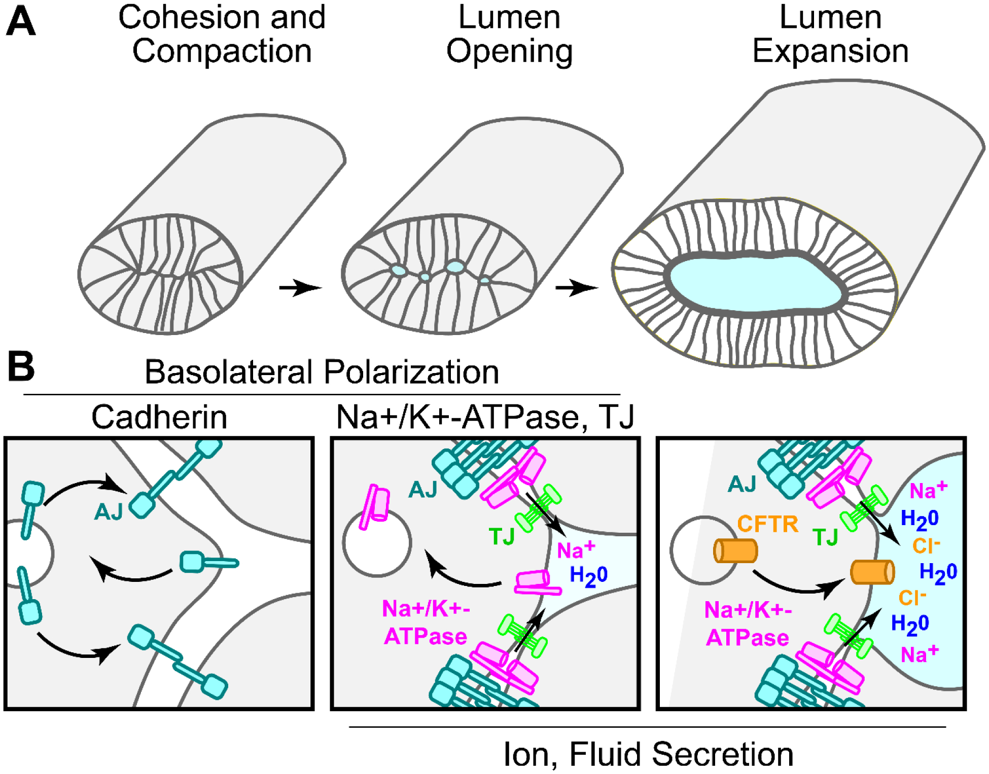
Fig. 1. Proposed mechanisms of lumen initiation and growth in vertebrate unpolarized epithelial organs.

(A) Basic morphogenetic events occurring during de novo lumen formation in newly polarizing organ precursors. Cell adhesion is a fundamental requirement that establishes positional domains required for proper development of other critical adhesion complexes. (B) Minimal molecular events that may facilitate lumen morphogenesis. Left panel: After differentiation, epithelial cells express adherens junction (AJ, cyan) proteins such as E-Cadherin that mediate cohesion. Transcellular interactions of cadherin extracellular domains stabilize the AJ at the newly forming basolateral domain. Cadherins lacking trans-interactions undergo internalization (inward arrow) and recycling (outward arrow). Middle panel: Cohesion and basolateral polarization facilitates transcellular interaction of Na+/K+-ATPase (magenta) ß-subunits, thereby stabilizing the complex. Like cadherins, Na+/K+-ATPase lacking trans-interactions are subject to internalization (inward arrow). Basolateral polarization also promotes the formation of tight junctions (TJ, green), allowing the epithelium to establish selectively permeable paracellular barriers to mediate ion and fluid transport. Hydrostatic pressure commences lumen opening. Right panel: Continued activity of ion channels such as basolateral Na+/K+-ATPase and apical CFTR in vertebrates drives enhanced fluid transport needed for lumen expansion.
