Fig. 1.
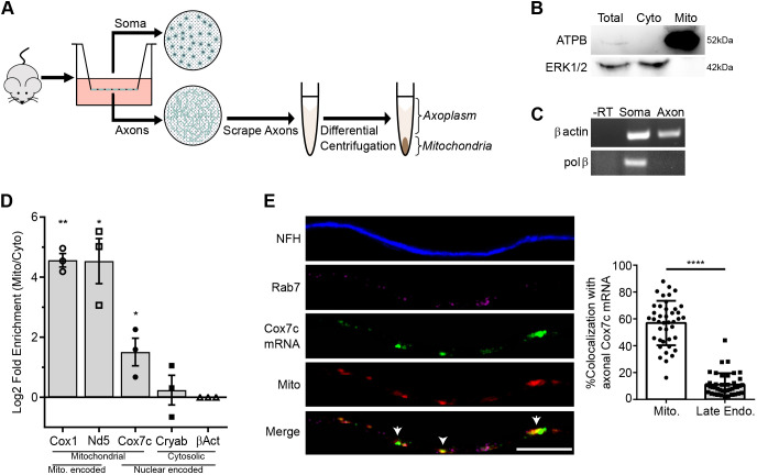
Nuclear encoded mitochondrial gene Cox7c mRNA is associated with mitochondria in motor neuron axons. (A) Schematic workflow used for membrane-based compartmental isolation of primary motor neuron axons. (B) Western blot analysis for fractionated axonal samples, with a mitochondrial marker (ATPB) and cytosolic marker (ERK1/2) demonstrating purity of mitochondria samples. (C) RT-PCR analysis for soma-specific mRNA (Pol β) confirming separation purity. β-actin represents mRNA present in both fractions. -RT, without reverse transcriptase. Images in B and C representative of three independent biological repeats. (D) RT-qPCR analysis was performed on axonal RNA samples from mitochondrial and axoplasm fractions for mRNAs encoded in the mitochondria (Cox1 and Nd5) or the nucleus (Cox7c, Cryab and β-actin). All values are normalized to β-actin transcript levels. Error bars are s.e.m.; n=3 independent biological repeats. *P<0.05, **P<0.01 (two-way ANOVA with Holm–Sidak correction). (E) Left, representative images of smFISH performed on primary motor neurons for Cox7c mRNA (green) along with immunostaining for axons by neurofilament heavy chain (NFH, blue), late endosomes (Rab7 marker, magenta) and mitochondria (MitoTracker staining, red). Arrows indicate areas of colocalization between mitochondria and Cox7c mRNA. Scale bar: 10 µm. Right, colocalization analysis of Cox7c mRNA with mitochondria and late endosomes signals. Error bars are s.d.; n=41 axons from three repeats. ****P<0.0001 (unpaired two-sided t-test).

