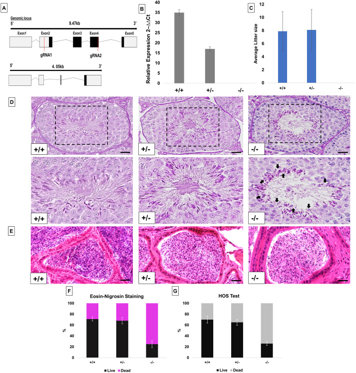
Fig. 1.
Generation and characterization of PFN4-deficient mice (Pfn4Δa). (A) Schematic of the Pfn4 locus and established PFN4-deficient mouse line. Red lines mark positions of the guide RNAs used for gene editing. (B) Validation of PFN4-deficient mice (Pfn4Δa). qRT-PCR was performed to check the relative expression of Pfn4 mRNA in murine testis of WT, Pfn4+/− and Pfn4−/− mice for the Pfn4Δa mutation. (C) Mating statistics of WT, Pfn4+/− and Pfn4−/− male mice (n=9/genotype). (D) PAS staining on WT, Pfn4+/− and Pfn4−/− testes sections. Dashed boxes indicate the areas shown at higher magnification below. Arrows indicate abnormal head morphology. (E) Hematoxylin and Eosin staining on WT, Pfn4+/− and Pfn4−/− cauda epididymis sections. (F) Quantification of E&N staining (n=3 biological replicates/genotype) of mature WT, Pfn4+/− and Pfn4−/− sperm isolated from cauda epididymis. (G) Hypo-osmotic swelling (HOS) test (n=3 biological replicates/genotype) of mature WT, Pfn4+/− and Pfn4−/− sperm isolated from cauda epididymis. Error bars represent mean±s.d. Scale bars: 10 μm.

