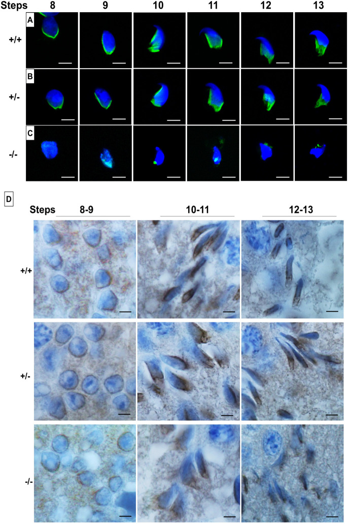
Fig. 3.
Manchette staining using α-tubulin and HOOK1 antibodies. (A-C) Immunofluorescence staining for the manchette using an α-tubulin antibody (green) on a germ cell population isolated from WT (A), Pfn4+/− (B) and Pfn4−/− (C) testes (n=3/genotype). Nuclei were stained with DAPI (blue). (D) IHC using an anti-HOOK1 antibody on WT, Pfn4+/− and Pfn4−/− testes sections. Scale bars: 20 µm.

