Fig. 5.
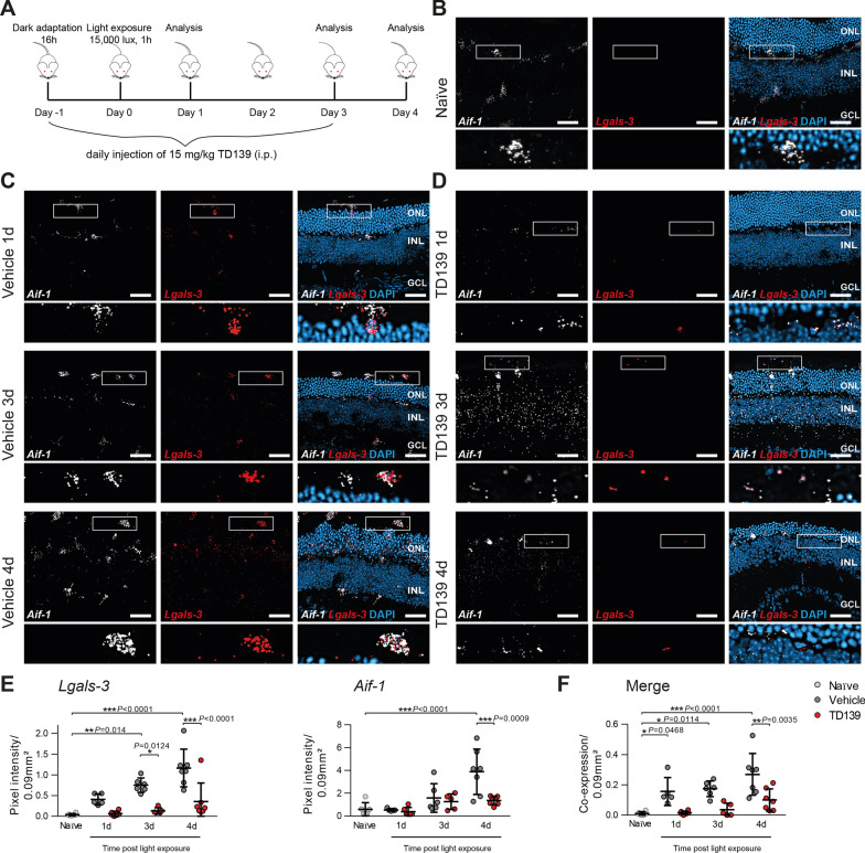
TD139 reduces Lgals-3 expression in light-exposed retinas. A Light exposure regimen of BALB/cJ mice and TD139 administration. B Representative images of in situ hybridization of retinal cross-sections from naïve BALB/cJ mice using mRNA probes for Aif-1 to label microglia/macrophages in combination with Lgals-3. Inlays show higher magnification. Scale bar: 50 µm. ONL, outer nuclear layer and INL, inner nuclear layer. C, D Representative images of in situ hybridization of retinal cross-sections of light-exposed mice treated with vehicle (C) or TD139 (D). Inlays show higher magnification. Scale bar: 50 µm. E, F Analysis of in situ hybridization signals of Aif-1 and Lgals-3 (E) and their merged co-expression (F) in light-exposed retinas at the indicated time points using pixel intensities within a defined area of 0.09 mm2 from the entire retina. Data show mean ± SEM. Naïve n = 6; Vehicle/TD139: 1d n = 5/6; 3d n = 6/5; 4d n = 7 retinal cross-sections. *P < 0.05 and ***P ≤ 0.001 by ordinary one-way ANOVA followed by Tukey’s multiple comparisons

