Figure 4.

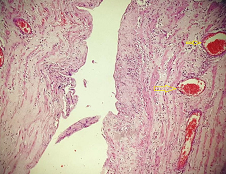
histological image shows that the muscular wall cyst is lined by a single layer of benign, flattened endothelium (arrows)

histological image shows that the muscular wall cyst is lined by a single layer of benign, flattened endothelium (arrows)