Fig. 4.
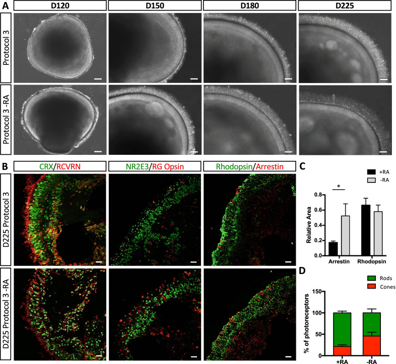
Qualitative and quantitative analysis of photoreceptor markers in the absence of RA. A Representative bright-field images of retinal organoids at D120, D150, D180 and D225 of differentiation using Protocol 3 (upper panels) or Protocol 3 without RA (Protocol 3 -RA; lower panels). Scale bars = 100 μm for D120 and 50 μm for D150, D180 and D225. B Representative IF images of D225 organoids generated using Protocol 3 (upper panels) or Protocol 3 -RA (lower panels): Left-hand panels, CRX (in green) and RCVRN (in red); middle panels, NR2E3 (in green) and RG opsin (in red); right-hand panels, rhodopsin (in green) and arrestin (in red). Scale bars = 20 μm. C Quantification analysis of the relative areas of arrestin and rhodopsin fluorescence within the ONL normalised to the area of Hoechst fluorescence in the ONL (see Additional file 1: Fig. S4). Quantification was performed on 3–5 images per organoid and 3 organoids were analysed per condition. Data are represented as mean ± SEM; *p < 0.05, n = 3; Mann and Whitney test. D Quantification of rods (green bar) and cones (red bar), as determined by areas of normalised rhodopsin and arrestin fluorescence, respectively, and expressed as a percentage of the total photoreceptors at D225 in Protocol 3 or Protocol 3 -RA organoids

