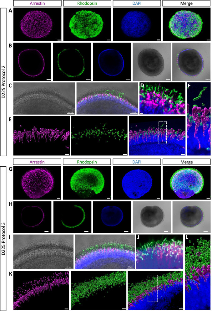
Fig. 6.
Three-dimensional imaging of mature retinal organoids. A Confocal imaging of D225 Protocol 2 organoids showing the arrestin-stained cones (in purple) and rhodopsin-stained rods (in green); DAPI-stained nuclei (in blue). Scale bars = 100 µm. B A single confocal plane showing the cones (in purple), rods (in green) and nuclei (in blue), the bright-field image, and a merge of the four channels. Scale bars = 150 µm. C Higher magnification of the bright-field and merged images in (B). Scale bars = 50 µm. D High resolution Aryscan imaging of the organoid surface showing the rods (in green) and cones (in purple); nuclei stained in blue. Scale bar = 10 µm. E Biphoton imaging and 3D reconstruction showing the cones (in purple), the OS-like segments of the rods (in green) and the merge of the two channels with the DAPI-stained ONL (in blue). Scale bars = 20 µm. Enlarged boxed area shown in (F). G Confocal imaging of Protocol 3 organoids showing cones (in purple) and rods (in green); DAPI-stained nuclei (in blue). Scale bars = 100 µm. H A single confocal plane showing the cones (in purple), rods (in green) and nuclei (in blue), the bright-field image and a merge of the four channels. Scale bars = 150 µm. I Higher magnification of the bright-field and merged images in (H). Scale bars = 50 µm. J High resolution Aryscan imaging showing the rods (in green) and cones (in purple); nuclei stained in blue. Scale bar = 10 µm. E Biphoton imaging and 3D reconstruction showing the cones (in purple), rods (in green) and the merge of the two with the DAPI-stained ONL (in blue). Scale bars = 20 µm Enlarged boxed area shown in (L)

