
Fig. 6.
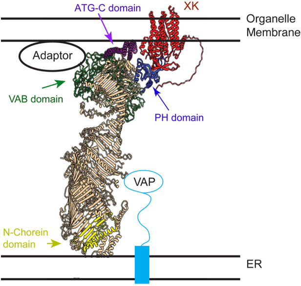
Model of VPS13A arrangement at a membrane contact site. A model structure of the VPS13A–XK complex based on assembly of Alphafold predictions is shown relative to the predicted locations of the ER membrane and a second organellar membrane (e.g. lipid droplet, mitochondria or plasma membrane). The XK protein is in red and the different domains of VPS13A are highlighted: yellow for the N-Chorein domain, green for the Vps13 adaptor binding (VAB) domain, purple for the ATG-C domain and blue for the PH-like domain. Binding to the VAP protein anchors the C-terminal end of VPS13A to the ER (Yeshaw et al., 2019).