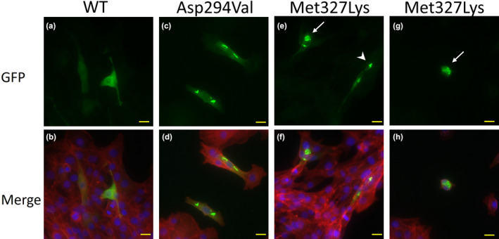
FIGURE 1.

WT and mutant ACTA1 proteins were expressed in C2C12 cells. The nucleus is stained by Fluoro‐KEEPER Antifade Reagent Nonhardening Type with DAPI (Nacalai Tesque), and F‐actin is stained by ActinRed™555 (Invitrogen). pAcGFP‐N1 plasmids with ACTA1‐WT, ACTA1‐Asp294Val, and ACTA1‐Met327Lys are transfected into C2C12 cells. The yellow bar indicates 20 μm. WT ACTA1 is expressed in the nucleus and cytoplasm with few aggregations (a, b). Asp294Val mutation (previously reported), positive control, shows needle‐like structures in the cytoplasm (c, d). Met327Lys mutation, as found in our case here, shows few aggregations in the cytoplasm and numerous apoptotic cells (e–h). White arrows indicate apoptotic cells (e, g), and the arrowhead indicates aggregations (e).
