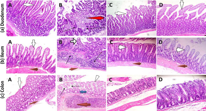
Figure 4.
Photomicrographs of the duodenum, ileum and colon in rats treated with cadmium chloride and Nigella sativa oil (H&E). A, control group; B, Cd only group; C, Cd+NSO group; D, NSO only group. (a) Duodenum: Villi (white arrows) morphology was largely normal in control, Cd+NSO and NSO groups; focal areas of ulceration (red arrow) and inflammatory cell infiltration (black arrows). (b) Ileum: Normal villi (white arrows) in all the groups, except the Cd group, which showed distorted villi arrangement and inflammatory cell infiltration (black arrow) in the sub-mucosal layer. Muscularis layer (red arrows) appears normal in all the groups (c) Colon: epithelium appears largely normal in the control, Cd+NSO and NSO groups. Lymphocytic infiltration (black arrows) and several lymphoid aggregates (red arrow) in the sub- mucosal layer

