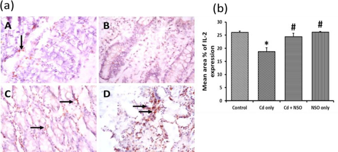
Figure 8.
(a) Immuno-histochemical staining of interleukin-2 (IL-2) in colonic sections of rats treated with cadmium and Nigella sativa oil X400; IL-2 expression is indicated as golden brown coloration (black arrows) on tissue structures. A, control; B, Cd only; C, Cd+NSO; D, NSO only. (b) The mean area% of IL-2 expression in the different groups. *indicates significant differences (p=0.032) compared to the control group; #indicates significant differences (p=0.025 and 0.041 for the Cd+NSO and NSO groups, respectively) compared to the Cd only group

