FIG. 2.
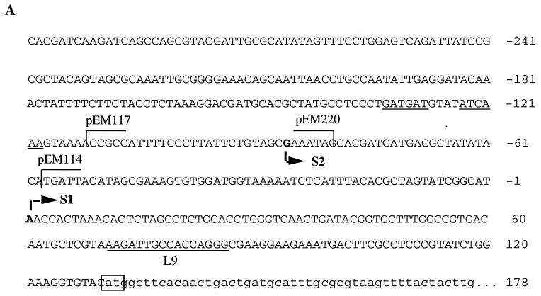
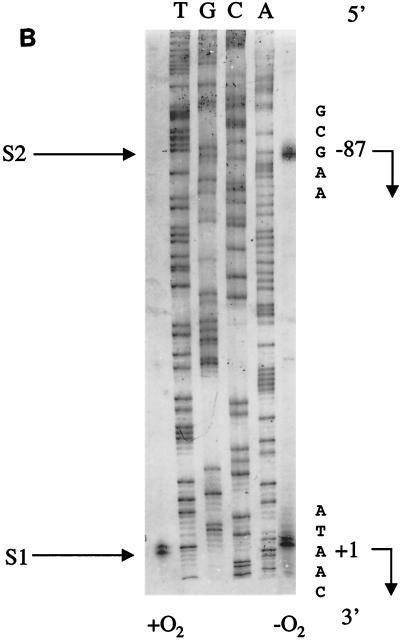
(A) Nucleotide sequence of the moa promoter region. Part of the moa upstream sequence is shown. Indicated are the different plasmid-borne 5′ (pEM114 and pEM117) and 3′ (pEM220) deletion end points. The position of binding of the primer (L9) used in the primer extension reactions is indicated by underlining and the L9 label. Transcriptional start sites, termed S1 (proximal) and S2 (distal), are shown in bold type. The Fnr recognition half-sites are underlined. The start codon of moaA is boxed and is in lowercase type, as is the portion of the moaA sequence shown. (B) Primer extension analysis of moa transcription. RNA was recovered from parental (RK4353) cells carrying plasmid pEM101, grown both aerobically (+O2) and anaerobically (−O2) in LB medium. The autoradiograph was reversed prior to photography to show the sequence of the coding strand. The DNA sequence shown was obtained using primer L9 (see Materials and Methods). The nucleotide sequence around each initiation site is shown on the right. The transcript labeled as S1 begins at nucleotide +1, and transcript S2 begins at position −87.


