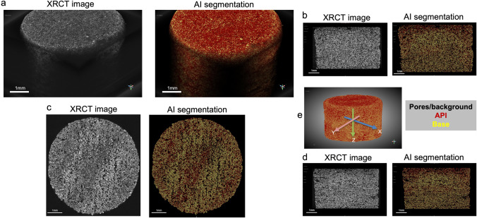
Fig. 7.
3D reconstructions of hydroxychloroquine sulfate 3D printed tablet of original XRCT images: black, pores or background; dark-gray, hydroxychloroquine sulfate; bright-gray, calcium sulfate hemihydrate (Left) and AI segmented images: black, pores or background; red, hydroxychloroquine sulfate; yellow, calcium sulfate hemihydrate (Right). Scale bars: 1 mm. (b) XZ cross-section images of hydroxychloroquine sulfate 3D printed tablet. (c) XY cross-section images of hydroxychloroquine sulfate 3D printed tablet. (d) YZ cross-section images of hydroxychloroquine sulfate 3D printed tablet. (e) Illustration of the 3D AI reconstructed tablets in X, Y, and Z axis.

