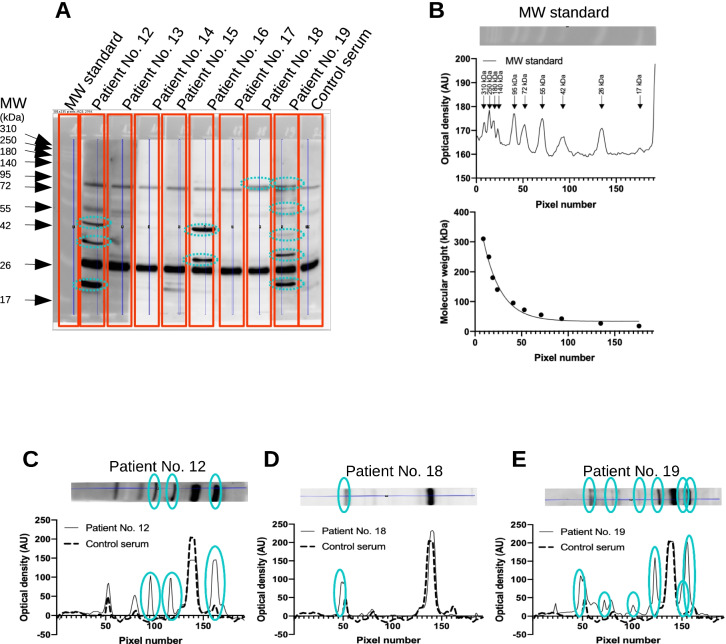
Fig. 1.
Detection of anti-cardiac autoantibodies in the sera of COVID-19 patients. Human heart homogenate was separated by SDS-PAGE (10% discontinuous gels, 80 µg proteinwell). A pre-stained standard was used to estimate molecular sizes (MW standard, panels A and B). Membranes were cut to strips after blocking and incubated separately with human serum samples (indicated in panel A) at a dilution of 1:1000. Strips were then fitted together and bound human IgG and IgM (shown in panel A) was detected by peroxidase labelled secondary antibodies. Densitometry was performed on the recorded images by ImageJ software, to yield density plots (shown in panels B–E by dashed (control serum) or continuous (COVID-19 patients’ sera) lines). Pre-stained standard proteins were used to construct a calibration curve (panel B lower graph), and molecular size of autoantigens was interpolated according to this calibration curve by GraphPad Prism software. Bands not apparent on the strip incubated by control serum were considered to represent anti-cardiac autoantibody binding to cardiac antigens and are indicated in panels C–E

