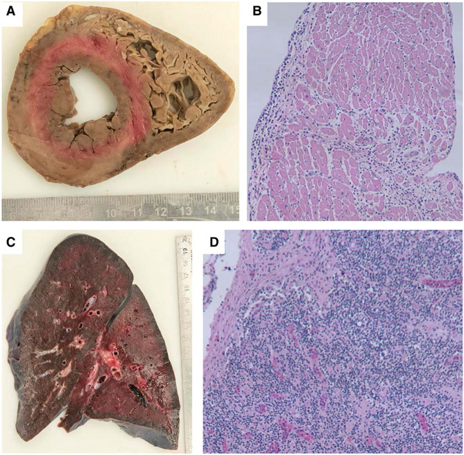
FIGURE 2.
Biventricular cross-section of heart (A) with areas of pallor. Cardiac myocytes with interstitial lymphoplasmacytic infiltrate (B, H&E stain, 100x). Mixed inflammation surrounding and involving a cardiac vessel (C, H&E stain, 100x). Focal area of myocyte damage in the right ventricle (D, H&E stain, 100x).

