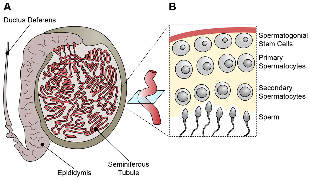
Figure 1.

Anatomy of the spermatogonial stem cell (SSC) niche.
(A) Human testes contain a web of seminiferous tubules, which are the site of spermatogenesis. They connect to the epididymis and the ductus deferens (or vas deferens) as a portal to ejaculation.
(B) Cross-section of a seminiferous tubule. SSCs proliferate and self-renew, producing spermatocytes, which undergo meiosis and, depending on their progression, are distinguished as primary or secondary. Following secondary meiotic division, spermatids differentiate into mature sperm (or spermatozoa) that will shed into the lumen of the seminiferous tubule prior to ejaculation.
