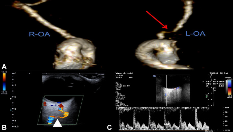
Figure 1.
Diagnostic workup for case 002. (A) 3T brain MR angiography 3D reconstruction depicting the origin of both ophthalmic arteries (OAs). The red arrow denotes stenosis in the long limb segment of the ophthalmic artery. EcoDoppler analysis, white (B) the arrow head depicts stenotic segment of the ophthalmic artery with (C) peak systolic velocities in the flow velocity waveform.

