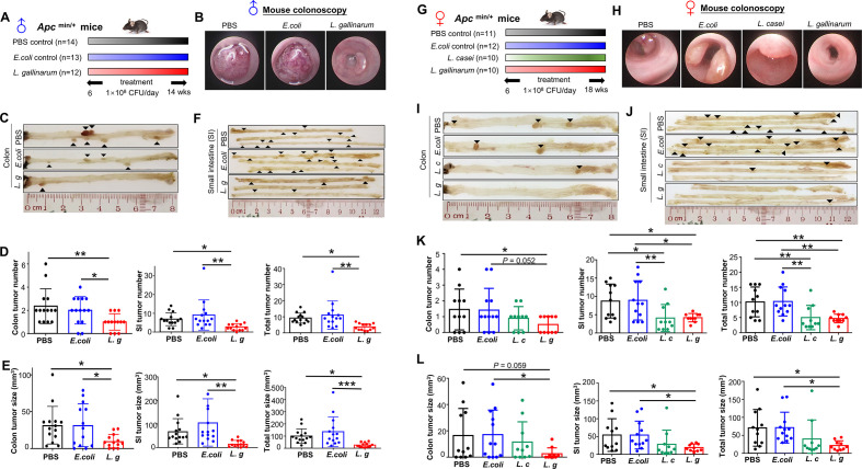
Figure 1.
L. gallinarum protects against intestinal tumourigenesis in Apc Min/+ mice. (A) Schematic diagram showing the experimental design, timeline of male Apc Min/+ mouse model. (B) Representative images of colon tumour from male Apc Min/+ mouse model. Colonoscopy confirmed that colon tumour size in L. gallinarum group was visually smaller than tumours in Escherichia coli MG1655 or PBS control groups. In both E. coli MG1655 and PBS control groups, colonoscope could not pass through the tumours. (C) Representative macroscopic images of colons from male Apc Min/+ mouse model. (D) Colon, small intestinal and total tumour number (colon+small intestinal) in male Apc Min/+ mice under different treatments. (E) Colon, small intestinal and total tumour size (colon+small intestinal) in male Apc Min/+ mice under different treatments. (F) Representative macroscopic images of small intestines from male Apc Min/+ mouse model. (G) Schematic diagram showing the experimental design, timeline of female Apc Min/+ mouse model. (H) Representative colonoscopic images of colon tumour from female Apc Min/+ mouse model. (I) Representative macroscopic images of colons from female Apc Min/+ mouse model. (J) Representative macroscopic images of small intestines from female Apc Min/+ mouse model. (K) Colon, small intestinal and total tumour number (colon+small intestinal) in female Apc Min/+ mice under different treatments. (L) Colon, small intestinal and total tumour size (colon+small intestinal) in female Apc Min/+ mice under different treatments. Each black triangle indicates one tumour location. P values are calculated by one-way analysis of variance. *P<0.05, **p<0.01, ***p<0.001. L. c, Lactobacillus casei; L. g, Lactobacillus gallinarum; PBS, phosphate-buffered saline; SI, small intestine.

