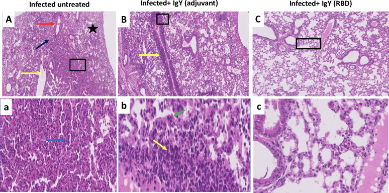
Fig 6.
(A and a) Histopathology of the lungs of Ad5-hACE2-transduced mice on day 6 after infection with SARS-CoV-2, showing characteristic bronchioalveolar interstitial pneumonitis with marked alveolar changes represented by alveolar wall thickening due to mononuclear cells infiltration, alveolar pneumocytes type 2 hypertrophic changes and occasionally associated alveolar wall hyalinization (blue arrow). Large number of the alveoli was involved with complete or partial obstruction and presence of some emphysematous alveoli (black star). Some bronchioles appear dilated with presence of exudative fluid in their lumina (yellow arrow). Some blood vessels show partial destruction of their walls with fluid and erythrocytic agglutination (red arrow). (B and b) a perivascular delayed hypersensitive lympho-plasmacytic aggregates with partial vascular wall destruction (yellow arrow) together with moderate alveolitis and alveolar wall thickening and or hyalinization due to pneumocytes hypertrophy and activated alveolar macrophages (green arrow). Some alveoli showed intraluminal aggregates of nonnuclear cells. (C and c), showing apparently normal lung tissue with preserved healthy bronchial tree, alveolar ducts, alveoli besides vascular and stromal structures. Scale bars: 200μm (upper row) and 50μm (lower row).

