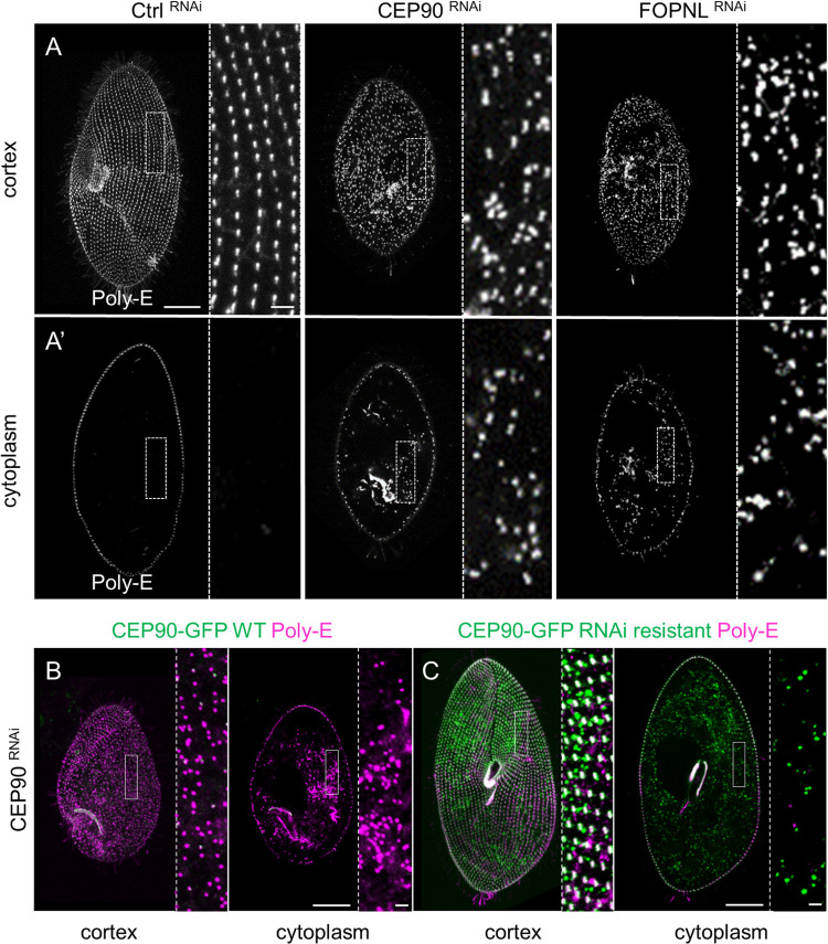
Fig 2. CEP90 depletion affects basal body docking.
(A, A’) Projection of confocal sections through the ventral surface (A) or at the intracytoplasmic level (A’), labeled with poly-E antibodies (grey) in control (Ctrl) RNAi, CEP90RNAi and FOPNLRNAi Paramecium. In control-depleted cells, BB are organized in parallel longitudinal rows highlighted in the magnification (right panel) (A) with no BB observed in the cytoplasm (A’). Cells observed after 2 divisions upon CEP90 or FOPNL depletion appear smaller than control ones, with a severe disorganization of the BB pattern (A). Numerous internal BB are observed in the cytoplasm (A’). Scale bars = 20 μm and 2 μm (magnification). (B, C) Specificity of CEP90RNAi: transformants expressing either WT CEP90-GFP (B) or RNAi-resistant CEP90-GFP (C) were observed after 2–3 divisions upon CEP90 depletion and analyzed for green emitted fluorescence and BB staining using poly-E antibodies. Scale bars = 20μm and 2 μm (magnification). The green fluorescence of WT CEP90-GFP transformants was severely reduced along with the expected disorganization of BB pattern at the cell surface (B). In contrast, the expression of RNAi-resistant CEP90-GFP rescued endogenous CEP90 depletion and restored the cortical organization of BB (C). BB, basal body; RNAi, RNA interference; WT, wild-type.

