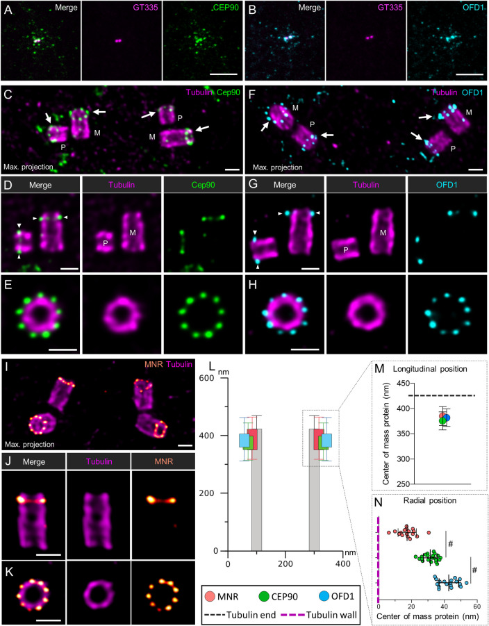
Fig 5. Centrosomal localization of CEP90 and OFD1 in mammalian cells.
(A, B) RPE1 cells were stained with anti-GT335 antibody directed against tubulin polyglutamylation (magenta) and anti-CEP90 (green) (A) or anti-OFD1 (cyan) (B). Both CEP90 and OFD1 decorated the centrosome and the centriolar satellites. Scale bar = 5 μm. (C-J) U2OS cells were expanded using the U-ExM technique and stained for tubulin (magenta) and CEP90 (green, C-E), OFD1 (cyan, F-H), or MNR (orange/red, I-J). Maximum intensity projections show the organization of CEP90 (C), OFD1 (F), and MNR (I) at the level of the centrosome in duplicating cells (M: Mature centriole; P: Procentriole). Single plan images show that CEP90 (D), OFD1 (G), and MNR (J) localize slightly underneath the distal end of both the mother (M) as well as the procentrioles (P). Finally, images of top-viewed oriented centrioles show 9-fold symmetry organization of CEP90 (E), OFD1 (H), and MNR (K) at the distal end of the centriole. Scale bars = 200 nm. (L, M) Distance between the proximal part of centriole (tubulin) and the fluorescence center of mass of CEP90 (green), OFD1 (cyan), or MNR (orange) (longitudinal position). N = 23, 25, and 49 centrioles for CEP90, OFD1, and MNR, respectively, from 3 independent experiments. Average +/− SD: CEP90 = 375.3 +/− 17.8 nm, OFD1 = 381.4 +/− 17.4 nm, MNR = 385.1 +/− 18.7. (L, N) Localization of CEP90, OFD1, and MNR with respect to the microtubule wall (radial position). Note that MNR localized the closest at the external surface of the microtubule wall, while OFD1 localizes more externally than CEP90. N = 21, 18, and 17 centrioles for CEP90, OFD1, and MNR, respectively, from 3 independent experiments. Average +/− SD: MNR = 17.4.1 +/− 5.5; CEP90 = 31.4 +/− 4.3 nm, OFD1 = 42.7 +/− 5.9 nm. Statistical significance was assessed by Kruskal–Wallis test followed by Dunn’s post hoc test, CEP90 vs. MNR p = 0.06 (#), CEP90 vs. OFD1 p = 0.07 (#). Source data can be found in S3 Data. MNR, Moonraker; U-ExM, ultrastructure expansion microscopy.

