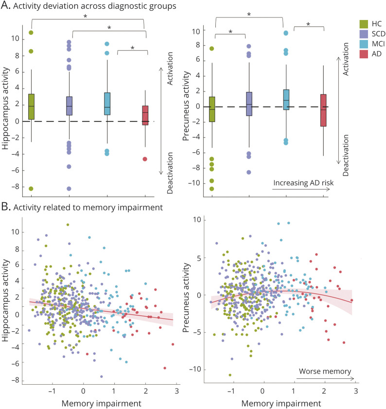
Figure 1. Differences in Region-Specific Novelty Activity Between Diagnostic Groups and With Increasing Memory Impairment.
(A) Mean fMRI activity (raw betas) for the novelty contrast (novel—familiar scenes) in the hippocampus and precuneus across diagnostic groups. Hippocampal activity was reduced in AD relative to all other groups. Precuneus activity followed an inverted U-shaped pattern with more advanced risk stages for AD. *Significant group differences surviving Bonferroni-Holm correction for the 5 group comparisons of interest (AD < HC < SCD/MCI) with p < 0.05. (B) Activation deviations related to memory performance as a continuous measure of clinical impairment. The memory factor score was inverted (*−1) to represent memory impairment for display purposes. The hippocampus showed a linear but the precuneus a quadratic pattern of activity deviations with increasing memory impairment. AD = Alzheimer disease dementia; HC = healthy control; MCI = mild cognitive impairment; SCD = subjective cognitive decline.

