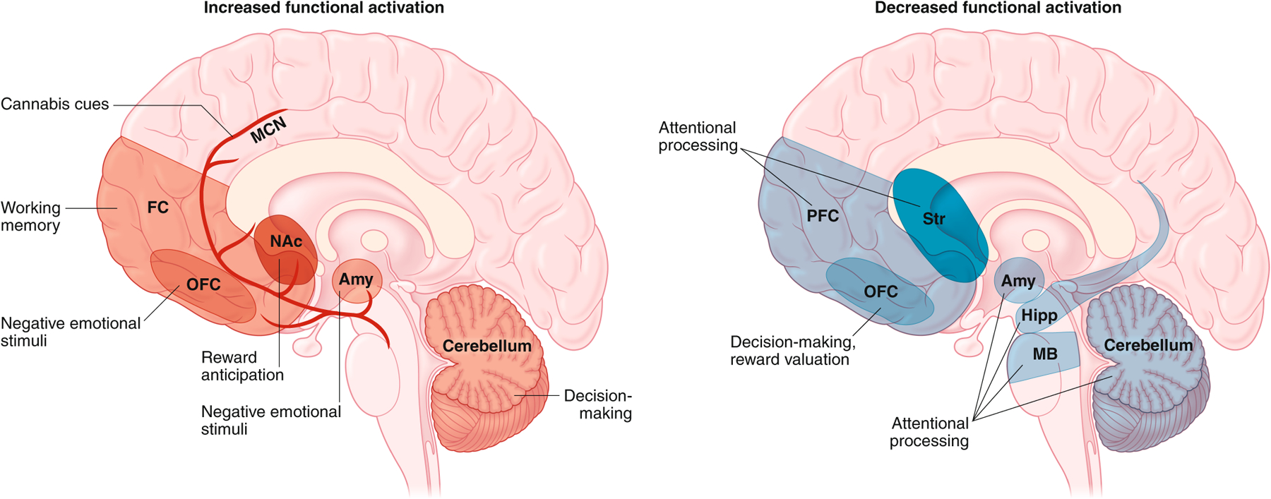
Fig. 4 |.

Differences in functional activity (based on functional MRI and electroencephalogram studies) detected in the brain of abstinent individuals with CUD during exposure to specific tasks and stimuli. Increased activation in specific regions is indicated in the left panel in red, and reduced activation is depicted in the right panel in blue. The specific task- or stimulus-driven alteration in activity is indicated in each region and circuit. Amy, amygdala; hipp, hippocampus; MCN, mesocorticolimbic network; FC, frontal cortex; NAc, nucleus accumbens; Str, striatum; MB, midbrain. Figures: Debbie Maizels/Springer Nature.
