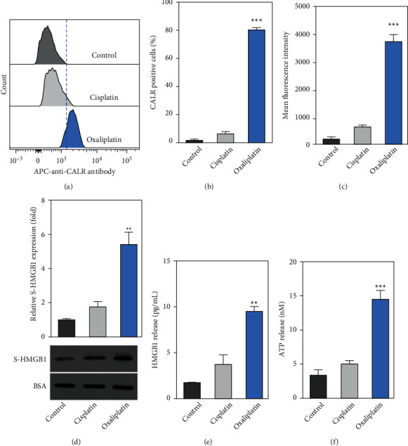
Figure 2.

Oxaliplatin induced immunogenic cell death (ICD) in Hep-2 cells. (a) After the cells were incubated with cisplatin (7.5 μM) or oxaliplatin (7.5 μM) for 24 h the cells were then stained with the APC-labeled anti-CALR antibody. The levels of surface calreticulin (CALR) in viable cells (refers to as PI negative) were determined by a flow cytometer. (b-c) The percentage of CALR-positive cells and the mean fluorescence intensity (MFI) were determined by flow cytometry. (d-e) Hep-2 cells were incubated with cisplatin or oxaliplatin. Next, the levels of HMGB1 in cell supernatant were determined by Western blotting and ELISA, respectively. (f) ATP released was determined by a commercialized kit. Data were represented as the means ± SD. ∗∗p < 0.01, ∗∗∗p < 0.001 compared with the cisplatin group.
