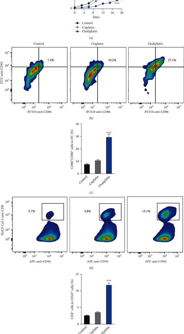
Figure 5.

In vivo antitumor effect of cisplatin and oxaliplatin in a mouse model of head and neck tumor. (a) Inhibition of tumor growth by various treatments on C3H/HeJ mice bearing murine mouse head and neck carcinoma SSC7 (n = 6). When tumor volumes reached about 50 mm3, mice received PBS, cisplatin, or oxaliplatin every three days for 4 times, the administration dosage of cisplatin and oxaliplatin was 3.0 mg/kg (i.v.). Data were represented as the means ± SD. ∗p < 0.05 (versus cisplatin group). (b-c) Flow cytometer gating and histogram analysis of matured DCs in the tumor tissues at the end of treatment (n = 6). The matured DCs were denoted as CD80+ CD86+ populations (gate in CD45+ CD11b+ CD11c + cell population). (d-e) Flow cytometer gating and histogram analysis of cytotoxic T cells (CD8+ T cells) in the CD45+ tumor-infiltrating immune cells in tumor tissues from mice receiving indicated treatment (n = 6). ∗∗∗p < 0.001 compared with the cisplatin group.
