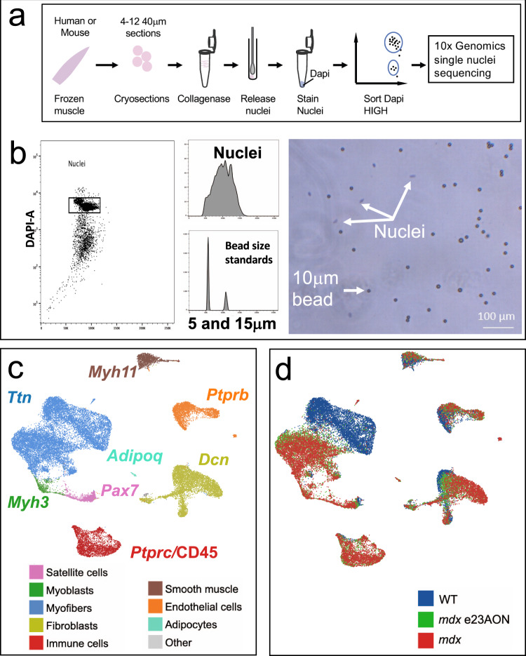
Fig. 1. Single nuclei sequencing identifies cell types and disease-related transcriptomic changes in murine muscle.
a Visual depiction of nuclei purification for snRNA sequencing. b Representative flow cytometric sorting of nuclei gated on DAPI staining (left panel), demonstration of forward scatter comparison to beads (middle panels), and size and morphology confirmation by visual microscopy (right panel). c Major clusters of cell types found in muscle were identified with a U-map analysis and subsequent identification of cell types using characteristic well known marker genes. One marker gene is shown for each cluster and text is colored to match the legend identifying major cell types of murine muscle. Subgroups with fewer members are listed in Other cell types panel, and include neural and schwann populations (gray). d Individual nuclei organized as the U-map identified clusters of c are labeled from mouse type: WT (blue), mdx (red), and mdx treated with e23AON (green).

