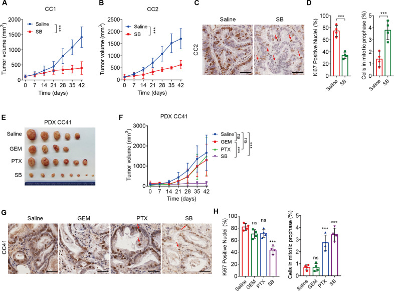
Fig. 6. Tumor suppression resulting from SB743921 treatment in CCA PDX models.
A, B Tumor volumes of CC1 and CC2 PDX models. PDX tumor-bearing mice were divided into two groups and treated with SB743921 (5 mg/kg, n = 4 and 3, respectively), or saline (n = 5 and 3 respectively). C, D Representative images and quantification of Ki67 staining in CC2 PDX tumors. Arrows indicate mitotic cells and the quantification of mitotic cells was shown. E Gross images of CC41 PDX tumors. F Tumor volumes of CC41 PDX models. PDX tumor-bearing mice were divided into four groups and treated with gemcitabine (10 mg/kg, n = 4), paclitaxel (10 mg/kg, n = 6), SB743921 (5 mg/kg, n = 8), or saline (n = 5). G, H Representative images and quantification of Ki67 staining in CC41 PDX tumors. Arrows indicate mitotic cells and the quantification of mitotic cells was shown. Scale bars, 50 μm. Panels A, B, and F were analyzed by two-way ANOVA; Panels D and H were analyzed by the two-tailed unpaired t-test. **p < 0.01, ***p < 0.001, ns, not significant.

