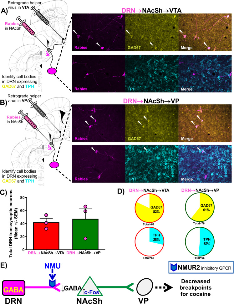
Fig. 4. GABAergic and serotonergic DRN → NAcSh neurons differentially innervated NAcSh → VTA and NAcSh → VP pathways.
A schematic diagram illustrates the viral placement with representative data in the DRN showing rabies (magenta) infected neurons that project onto the (A) NAcSh → VTA pathway or (B) NAcSh → VP pathway, colocalizing with GAD67 (yellow) or TPH (cyan). White arrows indicate colocalization events. C Total number of rabies positive neurons in the DRN from each rat in a survey of the DRN (15 coronal slices per rat and n = 3 per group). D Percentage of the rabies-positive neurons colocalizing with GAD67 or TPH. E The illustration summarizes the proposed pathway.

