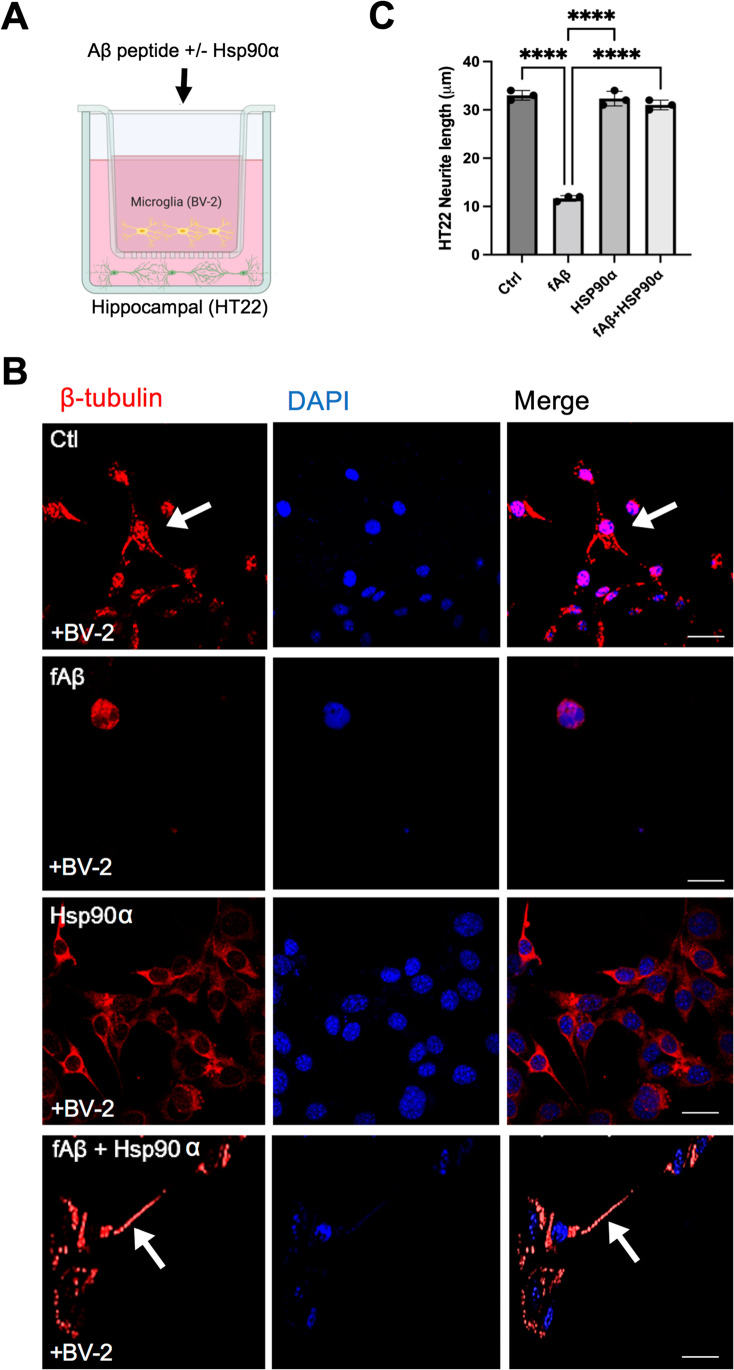
Fig. 4.
Extracellular Hsp90α mitigates fibrillar Aβ-induced neurotoxicity in vitro. A Schematic for BV2 microglia and HT22 hippocampal neuron co-culture. B HT22 hippocampal neuronal cells were grown on coverslips in the bottom layer of a transwell culture dish. BV2 cells were then added to the top layer of the transwell and incubated with no ligand (Ctrl) fibrillar f-Aβ1-42 (2 μM) (fAβ), Hsp90α (10 μg/ml), or f-Aβ1-42 + Hsp90α as indicated for 72 h. After the 72-h incubation, HT22 cells from the bottom wells were then fixed with 4% para formaldehyde and then permeabilized with 0.1% Triton X-100 before staining with anti-β-tubulin antibodies. Stained cells on coverslips were then examined by confocal microscopy. Scale bar = 5 μm. C β-tubulin-stained neurite outgrowth was measured using ImageJ. A total of 100 cells were counted in each sample. ****p < 0.0001 and n = 3. Cartoon created with BioRender.com. Experiments were repeated three times with similar results

