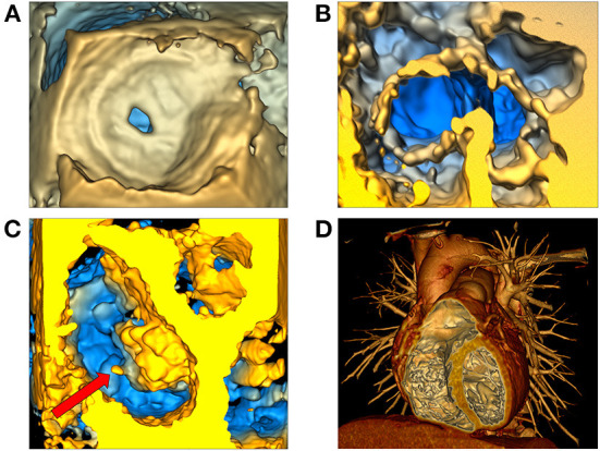
Figure 6.

Volume rendering of 3D echocardiographic and tomographic data. (A) Visualization of an atrial septal defect in the echo volume render module in SlicerHeart from a right atrial view; (B) Visualization of a complete common atrioventricular canal valve in diastole (ventricular view in SlicerHeart); (C) Left atrial view of an aortic valve and a mitral valve with anterior leaflet prolapse and a ruptured chord which is visible as indicated by the red arrow; (D) CT image of a normal anatomic heart cropped anteriorly to visualize the right atrium and ventricle and the left ventricle using volume rendering in 3D Slicer. CT, Computed Tomography.
