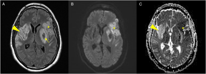
Figure 1.
Head imaging from Case 1. (A) Axial fluid inversion recovery MRI with (B) diffusion weighted imaging and (C) apparent diffusion coefficient images showing the chronic, evolving right MCA infarct from 2 months prior as demonstrated by gliotic changes on T2 FLAIR (arrowhead, A) and T2 shine-through effect on ADC (arrowhead, C) as well as the acute ischemic changes involving the left MCA territory (star, A-C) with area of hemorrhagic transformation (arrow, A).

