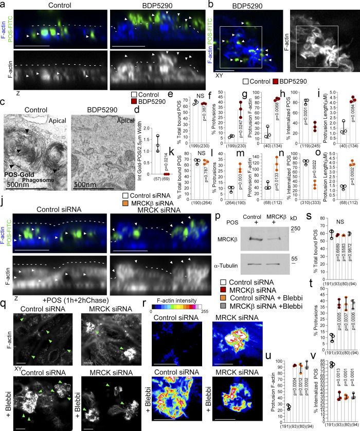
Figure 4.
MRCKβ and myosin-II control cup morphogenesis and POS internalization. (a–i) Inhibition of MRCKβ activity in primary porcine RPE cultures results in defective POS-induced apical membrane remodeling with stalled F-actin–based protrusions and inhibited phagocytosis as determined by confocal z-sections of porcine primary RPE cells stained with Atto647-Phalloidin; POS were labeled with FITC. White dashes highlight apical membrane and arrowheads internalized POS (left panels) or extracellular POS (right panels). TEM of primary porcine RPE cells using Gold-labeled POS confirmed MRCK kinase inhibition using BDP5290 results in an inhibition of phagocytosis. (j–p) Knockdown of MRCKβ resulted in similar defects in cup morphogenesis and phagocytosis. (q–v) Inhibition of myosin-II motor activity in MRCKβ-depleted cells results in identical defects in cup morphogenesis and phagocytosis, confirming that both defects are due to a single pathway. Note, heat maps signify F-actin intensity, white and green arrowheads highlight the position of POS, and all scale bars represent 10 μm. The quantifications in d–h are based on n = 3 independent experiments and show data points, means ±1SD (top of bar), and the total number of cells. P values were derived from t tests. Source data are available for this figure: SourceData F4.

