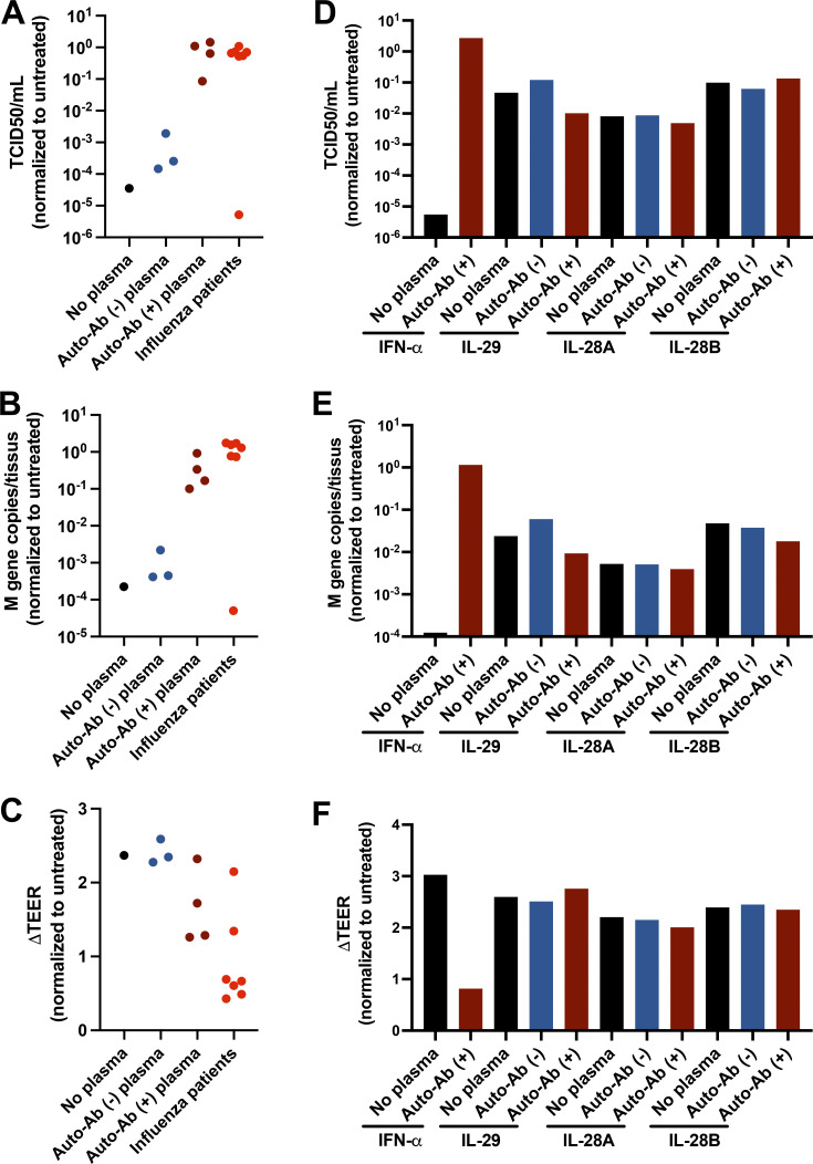
Figure 4.
Neutralizing auto-Abs block the antiviral function of IFN-α2 in IAV-infected HAE cultures. (A–F) HAE reconstituted from human nasal primary cells and maintained in an air–liquid interface were either left untreated or treated with 2 ng/ml exogenous IFN-α2a (A–C) or 20 ng/ml exogenous IL-29, IL-28A, or IL-28B (D–F), in the presence of inactivated patient plasma (1:100 diluted) for 24 h before IAV infection. Cells were treated again on the basolateral side with same concentration of IFN-α2a or IFN-λ1/IL-29, IFN-λ2/IL-28A, or IFN-λ3/IL-28B in the presence of patient plasma (n = 7) 1 h after IAV infection. These seven patients had auto-Abs neutralizing IFN-α at 10 ng/ml, but not IFN-β or -λ. HAE apical poles were washed, 54 h after infection, and titrated by TCID50 determination (A and D) and quantitative RT-PCR (B and E). Changes in TEER (ΔTEER) were measured as a surrogate for the integrity of HAE (C and F). Previously identified auto-Ab–positive (auto-Ab [+]) or auto-Ab [−] plasma samples were used as controls. Experiments were repeated three times.

