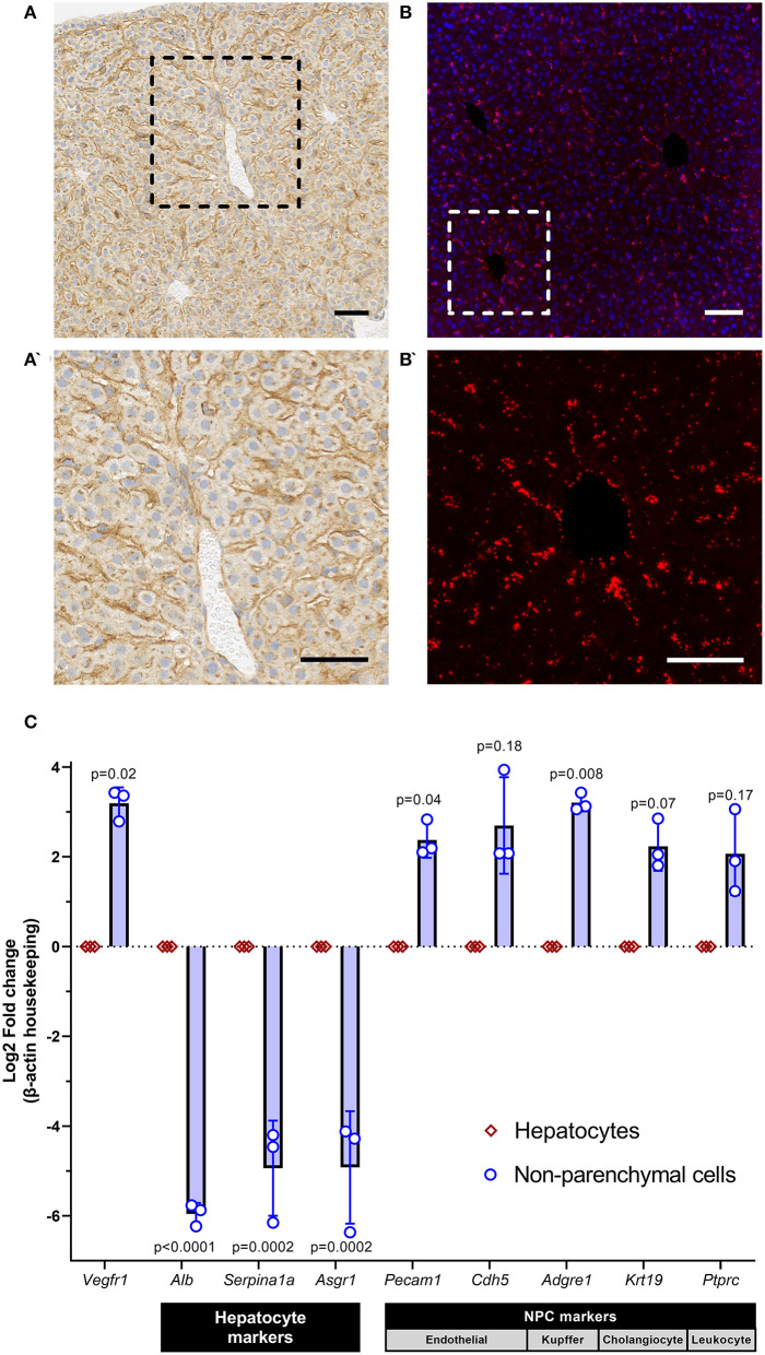
Figure 1.
sVEGFR1 is not significantly expressed by hepatocytes in the liver. (A,A') Immunohistochemistry of mouse liver demonstrates strong staining in the sinusoidal regions of the liver with negligible staining in hepatocytes; scale bar 50 μm. (B,B') RNAscope of mouse liver demonstrates hybridization signal in the sinusoidal regions of the liver; scale bar 50 μm. (C) Quantitative RT-PCR of isolated hepatocytes and non-parenchymal cells (NPCs) shows significantly greater expression of Vegfr1 in NPCs compared to hepatocytes; n = 3 wild-type mice and supporting gene expression patterns to confirm isolation of pure cell populations. Analysis with paired t-tests.

