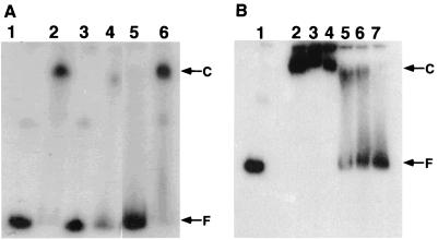
FIG. 3.
(A) Band shift assays of the CIRCE probe with B. thermoglucosidasius HrcA renatured with nonspecific DNA (pUC119). Positions of the radiolabeled free DNA probe (F) and HrcA-probe complex (C) are marked. B. thermoglucosidasius HrcA samples contained no urea. Lane 1, labeled CIRCE probe (0.2 μg) alone; lane 2, labeled CIRCE probe (0.2 μg) with renatured HrcA (0.1 μg); lane 3, labeled nonspecific probe (0.2 μg) alone; lane 4, labeled nonspecific probe (0.2 μg) with renatured HrcA (0.1 μg); lane 5, same as lane 2 with nonlabeled CIRCE probe (1 μg); lane 6, same as lane 2 with nonlabeled nonspecific probe (1 μg). (B) Thermostability analyses of HrcA and probe complex by band shift assays. The binding reaction of B. thermoglucosidasius HrcA to the CIRCE probe was done at room temperature for 10 min; then each mixture was incubated at the indicated temperature for 1 min. Immediately after heat shock treatment, the samples were loaded into wells of a polyacrylamide gel. Lane 1, labeled CIRCE probe (0.2 μg) alone; lanes 2 to 7, the labeled CIRCE probe (0.2 μg) with renatured HrcA (0.1 μg); lane 2, no heat shock treatment after the binding reaction (control); lanes 3 to 7, heat shock treatment at 40, 50, 60, 65, and 70°C, respectively. Positions of the radiolabeled free CIRCE probe (F) and HrcA-probe complex (C) are marked.

