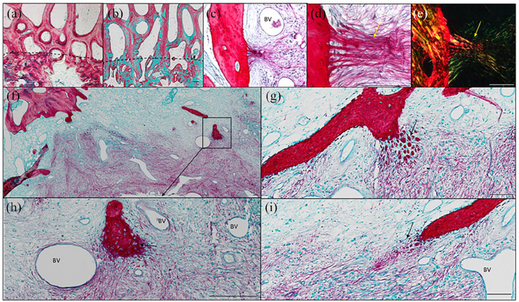
Figure 5.
Histological sections of Autograft group stained for Alcian Blue/Sirius red (a) and Goldners trichrome (b) depicting the integration boundary (dotted line) of autograft with native trabecular bone. Organized skeletal fibres (alcian blue/Sirius Red stain) extending from the bone (yellow arrows) into the implant of BMP2 + ACS + LLG (c and d). Polarized light microscopy depicting the organized collagen structures (e). Alcian blue/Sirius red staining of the matrix juxtaposed to the bone trabecular and implanted BMP2 + ACS + LLG in the condyle defect regions (f–i). Black arrows depict packets of new bone formation along with skeletal filaments (g and i), which are of a possible endochondral process (arrow) (h). BV = blood vessel. Scale bar = 100 µm.

