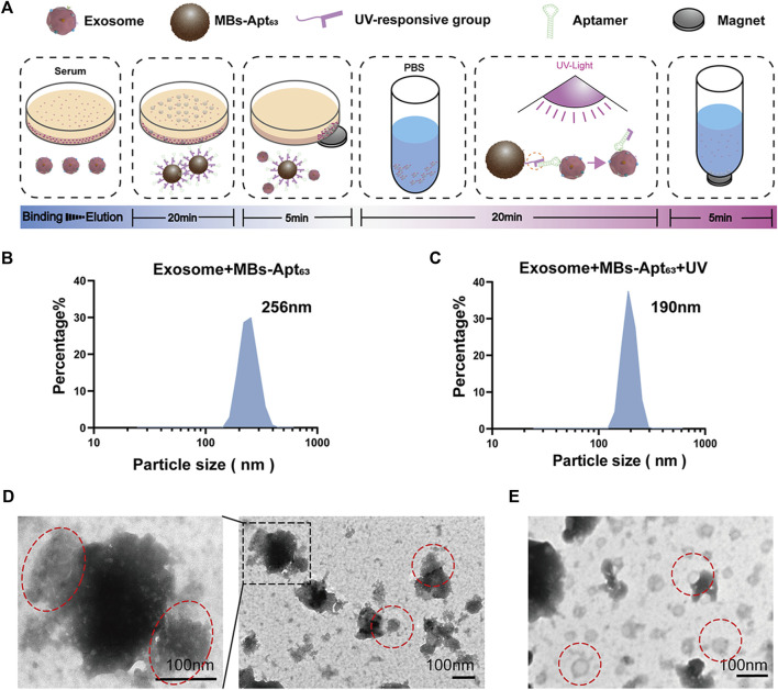
FIGURE 4.
Capturing exosomes by and elution of exosomes from MBs-Apt63. (A) Schematic diagram for extracting exosomes from serum and cell supernatant with MBs-Apt63; (B) DLS characterization after incubating exosomes with MBs-Apt63; (C) DLS characterization after exposing the MBs-exosome complex to UV light; (D) Transmission electron microscopy image of exosomes bound to MBs-Apt63 (left, 100000×, right, 25000×). Positions of exosomes and MBs-Apt63 are marked in red; (E) Transmission electron microscopy image of exosomes eluted from MBs-Apt63 by UV light.

