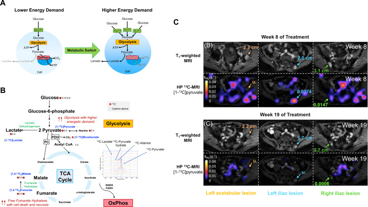
Figure 2.
Imaging glucose metabolism with HP 13C-MRI. (A) An increased expression of glucose transporters, for example, GLUT1 and elevated glycolysis is seen in cells with higher energy demand, for example, cancer cells and activated T cells. (B) Several 13C-labeled imaging probes have been developed for evaluating different downstream processes of the glucose metabolism pathway. These include [1-13C]pyruvate for imaging the kinetics of pyruvate-to-lactate conversion,78 13C-labeled bicarbonate (H13CO3 −) for detecting tumor pH in vivo,79 and increased production of [1,4-13C2]malate from the administered [1,4-13C2]fumarate as a surrogate biomarker of cell necrosis or tumor cell death in response to treatment.80 (C) HP 13C-MRI of a patient with prostate cancer showed a marked decrease in [1-13C]pyruvate to [1-13C]lactate conversion, with a corresponding reduction in tumor size, in the three bone metastases (left acetabular, left and right iliac lesions) between week 8 and 19 following pembrolizumab treatment. Images adapted and reproduced with permission from de Kouchkovsky et al.87 (A) and (B) are graphics created by the author (DL). ATP, adenosine triphosphate; NADH, reduced nicotinamide adenine dinucleotide; FADH, reduced flavin adenine dinucleotide; TCA, tricarboxylic acid; PC, pyruvate carboxylase ; PDH, pyruvate dehydrogenase.

