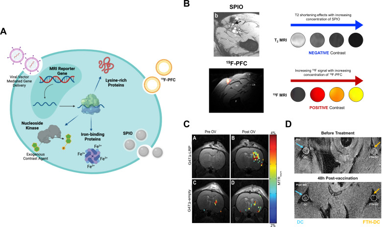
Figure 3.
MRI tracking of leukocytes and viral vector-mediated gene therapy. (A) Graphical overview of direct and indirect cell labeling approaches with superparamagnetic iron oxide (SPIO) nanoparticles, 19F-perfluorocarbons (PFC), and examples of MRI reporter genes. (B) A comparison between the negative contrast (decreased signal) obtained with SPIO versus the positive contrast (increased signal) obtained with 19F-PFC labeling of human dendritic cells. (C) Chemical exchange saturation transfer MRI of lysine-rich protein (LRP) concentration in rat glioma tumors before and at 8 hours following G47Δ oncolytic viral (OV) therapy showed higher signal on the magnetization transfer ratio asymmetry (MTRasym) maps of tumors injected with G47Δ-LRP but not the empty vector. (D) T2-weighted MRI showed signal loss or decreased T2 signal (orange arrow) at the popliteal lymph node near the footpad of a mouse at 48 hours following injection with dendritic cells expressing ferritin (FTH-DC). (A) is a graphic created by author (DL) using Biorender (publication licence AU24ELW1E5). Images in (B), (C) and (D) are reproduced with permission from Ahrens et al, Farrar et al, de Vries et al 110 123 170 and Kim et al.124

