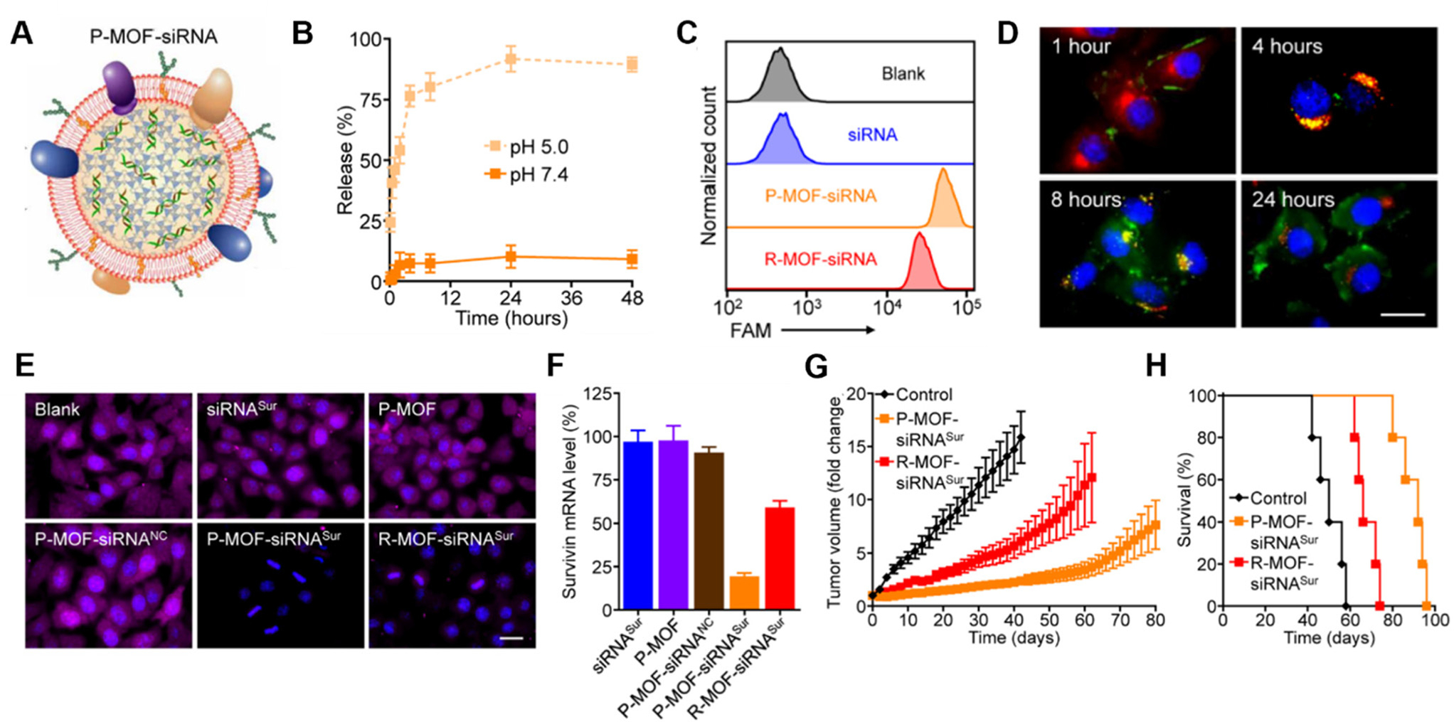
Fig. 4.

Representative example of a membrane-wrapped siRNA nanocarrier. (A) Platelet membrane-coated siRNA-loaded MOFs (P-MOF-siRNA) were generated by mixing the siRNA payload with Zn2+ and 2-methylimidazole (mim), followed by coating with a natural cell membrane derived from platelets. (B) siRNA release from P-MOF-siRNA at pH 5.0 or pH 7.4 over time (n = 3, mean ± SD). (C) Flow cytometry analysis of siRNA uptake in SK-BR-3 cells 24 hours after incubation with free siRNA, P-MOF-siRNA, or R-MOF-siRNA. (D) Fluorescence microscopy images of siRNA localization in SK-BR-3 cells 1, 4, 8, and 24 hours post incubation with P-MOF-siRNA (scale bar = 20 μm; siRNA = green; nuclei = blue; endosomes = red). (E) Immunofluorescence analysis of survivin protein expression in SK-BR-3 cells treated with different siRNA nanocarriers for 48 hours (scale bar = 20 μm; survivin = purple; nuclei = blue). (F) PCR analysis of relative survivin mRNA expression in SK-BR-3 cells after incubation with various siRNA nanocarriers for 48 hours (n = 3, mean + SD). (G) Growth kinetics of subcutaneous SK-BR-3 tumors in nu/nu mice treated intravenously with P-MOF-siRNASur or R-MOF-siRNASur every 3 days for four total administrations (n = 5; mean ± SEM). (H) Survival of the mice in (G) over time (n = 5). From ref. 41 (J. Zhuang, et al., Target gene silencing in vivo by platelet membrane-coated metal–organic framework nanoparticles, Sci. Adv., 2020, 6). Reprinted with permission from the American Association for the Advancement of Science (AAAS), copyright 2020.
