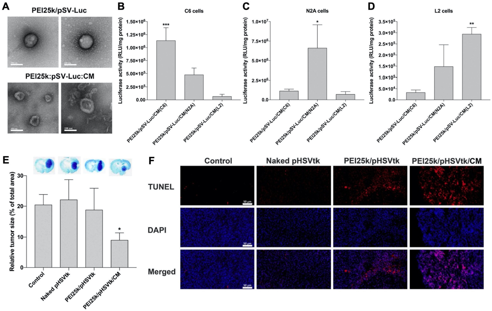
Fig. 5.

Representative example of a membrane-wrapped pDNA carrier. (A) TEM images of unwrapped PEI25k/pSV-Luc complexes and PEI25k/pSV-Luc/CM nanoparticles prepared at a 1 : 1 : 20 weight ratio. Scale bar indicates 100 nm. (B–D) Luciferase transfection efficiency of PEI25k/pSV-Luc/CM nanoparticles prepared with cell membranes from (B) C6, (C) N2A, and (D) L2 cells and transfected into different cell types to assess homotypic targeting and pDNA delivery. The data indicate the mean ± standard deviation of quadruplicated experiments. ***P < 0.001 compared with the other groups. *P < 0.05 compared with the other groups. **P < 0.01 compared with PEI25k/pSV-Luc/CM(C6), but no statistical significance compared with PEI25k/pSV-Luc/CM(N2A). (E) Analysis of tumor suppression enabled by pHSVtk delivery. The PEI25k/pHSVtk/CM nanoparticles and PEI25k/pHSVtk complexes were intratumorally injected into C6 glioblastoma tumors in rats. After 7 days, the brains were harvested and subjected to Nissl staining to quantify relative tumor size by measuring tumor areas in ImageJ. *P < 0.05 compared with the other groups (n = 6). (F) TUNEL assay of excised tumors from the study described in (E). Scale bar indicates 50 μm. Reproduced from ref. 55 (S. Han, et al., J. Controlled Release, 2021, 338) with permission from Elsevier BV, copyright 2021.
