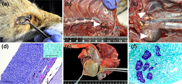
Fig. 1.
Red fox with generalized tuberculosis and canine distemper virus coinfection. Gross and histopathological findings. a Right eye, mild corneal edema and hypopyon, and few filiform worms consistent with Thelazia callipaeda in the conjunctival sac. b Cranial mediastinal lymph node with multifocal yellow areas of caseous necrosis. c Multiple foci of caseous-purulent necrosis in both kidneys. d Right eye, dense perivascular to diffuse infiltration of the choroid with histiocytes and lymphocytes, and a dense subretinal pyogranulomatous exudate (Hematoxylin and eosin stain, original magnification × 200, bar = 100 μm), Inset, pyogranulomatous exudate with multiple purple-stained acid-alcohol resistant bacteria (Ziehl-Neelsen stain, original magnification × 200, bar = 100 μm). e Diffuse thickening of the pericardium and abundant caseous-purulent exudate filling the pericardial cavity. f High quantity of acid-alcohol resistant bacteria within macrophages and necrotic areas in the kidneys (Ziehl-Neelsen stain, original magnification × 400, bar = 100 μm). All images taken by the authors

