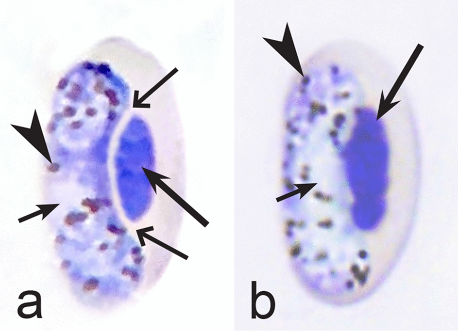
Fig. 9.

Morphological features of gametocytes, which are used for identification of Haemoproteus species parasitizing Cathartiformes birds. Macrogametocyte (a) and microgametocyte (b) of Haemoproteus catharti (a, b). Note that advanced growing gametocytes often do not adhere to erythrocyte nuclei (a). Pigment granules are of medium size and numerous (a, b). Long simple arrows—host cell nuclei. Short simple arrows—parasite nuclei. Simple arrowheads—pigment granules. Simple wide long arrows—unfilled space between growing gametocyte and nucleus of infected erythrocyte. Other explanations are given in the text
