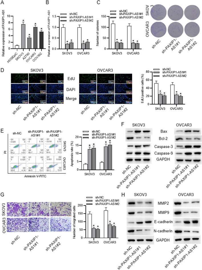
Fig. 1.
Expression pattern and functional role of PAXIP1-AS1 in OC cells. a RT-qPCR data of PAXIP1-AS1 expression in HOSEpiC cell line and OC cell lines. b Knockdown of PAXIP1-AS1 in SKOV3 and OVCAR3 cells validated by RT-qPCR. c-d Proliferation of SKOV3 and OVCAR3 cells upon PAXIP1-AS1 silencing was evaluated via colony formation assay and EdU assay. e Apoptosis of SKOV3 and OVCAR3 cells after PAXIP1-AS1 silencing was assessed through flow cytometry analysis. f Protein levels of Bax, Bcl-2, caspase-3 and caspase-9 under sh-PAXIP1-AS1 transfection were detected by western blot. g Migration of SKOV3 and OVCAR3 cells transfected with sh-PAXIP1-AS1 was confirmed by Transwell assay. h MMP2, MMP9, E-cadherin and N-cadherin protein levels were testified with western blot upon PAXIP1-AS1 knockdown. *p < 0.05

