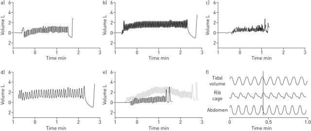
FIGURE 1.
Recordings of quiet tidal breathing at rest, followed by maximal expiration then inspiration. Breathing patterns are shown for: a) a healthy volunteer; b) hyperventilation syndrome, note the rapid respiratory rate, and tidal breathing closer to inspiratory capacity than in panel a; c) erratic breathing pattern, note that the patient was unable to coordinate a maximal expiratory and inspiratory manoeuvre at the end of the recording; d) thoracic dominant breathing, note the large volume breaths with minimal inspiratory reserve capacity; e) forced expiratory pattern before (grey) and after (black) exercise, note tidal breathing occurs at low lung volumes, with minimal expiratory reserve volume; and f) thoraco-abdominal asynchrony, this panel shows recordings from sensors detecting thoracic and abdominal movement, demonstrating asynchrony during quiet tidal breathing.

