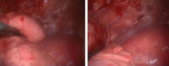
FIGURE 2.
Thoracoscopic images showing two different views of a mass enveloping the thoracic aorta and lesions connected to the thoracic wall, diagnosed as lymphangiomas at histological examination of the biopsy. Images courtesy of Spaggiari Lorenzo and Gasparri Roberto (Istituto Europeo di Oncologia, Milan, Italy).

