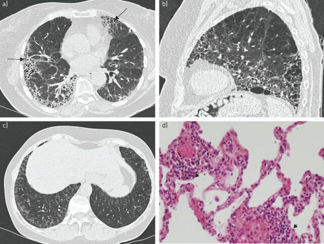
FIGURE 2.
a) Usual interstitial pneumonia in a 69-year-old woman with primary Sjögren's syndrome. High-resolution computed tomography (CT) showing bilateral reticular areas and honeycombing with peripheral and basal predominance (arrows). b) Combined pulmonary fibrosis and emphysema syndrome in a 68-year-old smoker with primary Sjögren's syndrome complicated by pulmonary hypertension. High-resolution CT shows bilateral reticular areas and honeycombing with posterior and basal predominance. Emphysema is predominant in apical areas. c, d) Lymphocytic interstitial pneumonia in a 59-year-old woman with primary Sjögren's syndrome and lymphocytic alveolitis. c) High-resolution CT shows thickening of interlobular septa with superimposition of intralobular reticulation. d) Photomicrograph (haematoxylin–eosin stain) shows diffuse thickening of alveolar septa and peribronchiolar infiltration with lymphocytes and plasma cells (arrowheads). Original magnification ×100.

