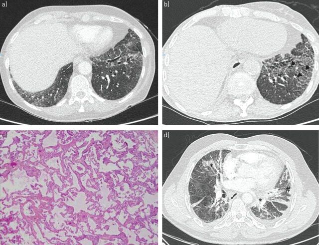
FIGURE 3.
Nonspecific interstitial pneumonia in a woman a, c) at the time of Sjögren's syndrome diagnosis and b) after 3 years. a, b) High-resolution computed tomography (CT) show bilateral areas of ground-glass attenuation and traction bronchiectasis (arrowheads) with peripheral intralobular reticulation. c) Photomicrograph (haematoxylin–eosin stain) shows diffuse and homogenous collagenous fibrosis in the alveolar area. d) Organising pneumonia and primary Sjögren's syndrome revealed by acute onset of pleuropneumonia in a 64-year-old man. High-resolution CT images show bilateral patchy areas of consolidation (#) and areas of ground-glass opacity (*). The patient was diagnosed with nonspecific interstitial pneumonia 2 years later.

