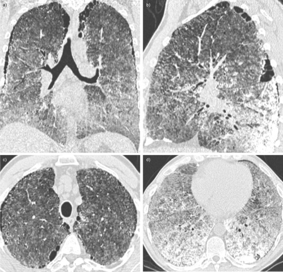
Figure 1.

Case 1. High-resolution computed tomography imaging of the lungs showing intra-alveolar calcifications, mainly subpleural, in posterior and basal parts of the lungs. Paraseptal emphysema is visible. a) Coronal view, b) sagittal view and c and d) axial view of the chest.
